HiPath FL
Unique Digital Pathology Workflow for Fluorescence Images
Illustrate with new image from Inova (Renal Pathology)

HiPath FL
in a nutshell
HiPath FL enables the scanning and visualization of fluorescence whole slide images at 20x or 40x magnification, supporting the review of fluorescent signals across the entire tissue section. This broader view supports a more accurate selection of relevant regions for high-magnification imaging and analysis.
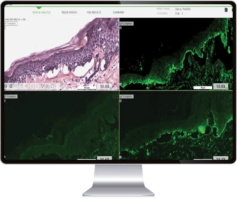
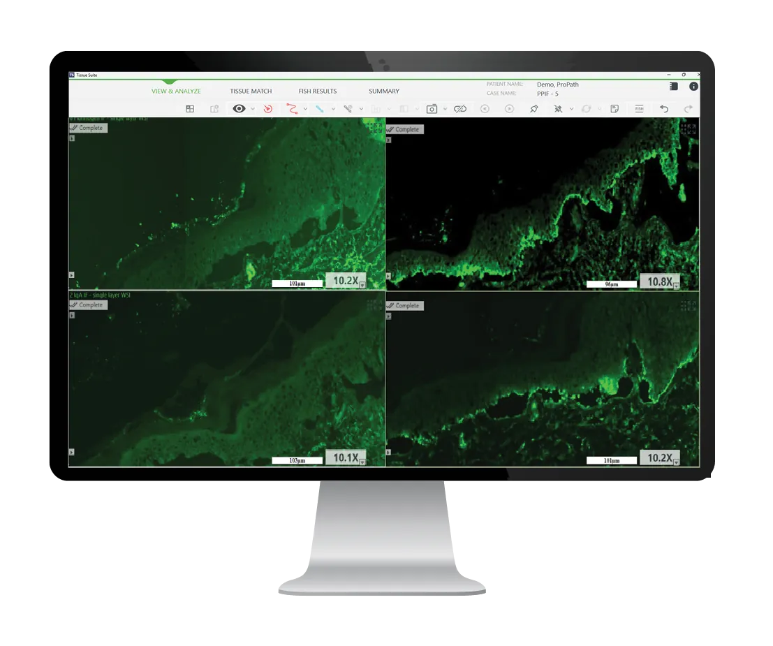
With its tissue matching capabilities, HiPath FL supports side-by-side visualization of up to four fluorescent specimens, enabling efficient review of FISH and immunofluorescence panels.
Experience the power of HiPath FL to deliver consistent, standardized results across both tissue FISH and immunofluorescence applications.
Benefits to your lab
Digital review of multiple fluorescent specimens side-by-side
Applicable to multiple tissue and cell types
Stain-vendor agnostic for enhanced lab flexibility
Archiving of entire fluorescent slide for future review and comparison
Digitalize Your Review of FISH and IF Staining Panels
HiPath FL streamlines immunofluorescence workflows by enabling the detection and evaluation of FITC-labeled biomarkers across multiple targets. Applicable to a wide range of tissue and cell types, including skin and kidney specimens, the platform provides a convenient and efficient environment for reviewing immunofluorescence staining panels such as IgG, IgA, IgM, C3, and other clinically relevant markers, helping to standardize interpretation and improve workflow efficiency.