

PathFusion
Bridging the Gap between Brightfield and Fluorescence Pathology


PathFusion
in a nutshell
PathFusion provides a comprehensive digital pathology workflow, seamlessly integrating brightfield and fluorescence imaging across multiple probe vendors and sample types. The application features state-of-the-art functionalities including computer-aided tissue detection, cell identification, signal detection, and cell classification.
The automatic selection of frames within the regions of interest marked on the H&E specimen, and transferred to the registered FISH slide, ensures more precise targeting for high-magnification tissue FISH scanning and analysis.
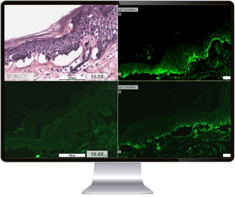
Furthermore, by enabling simultaneous display of up to four H&E and/or IF images side-by-side, PathFusion helps to conveniently correlate protein expression with morphological context.
With PathFusion, experience the transformative power of digital analysis of fluorescence tissue specimens.
Benefits to your lab
Unlimited Z-stacked layers and automatic detection of faint FISH signals
High Image Quality with automated optimization
and enhancement
Whole Slide Imaging
with intuitive viewing
and advanced tools
User Independent Scanning for increased productivity and greater slide volumes
Computer-Aided Tissue FISH Detection: Unlocking Quantitative FISH Analysis
With ASI’s proprietary algorithms, PathFusion delivers unparalleled image quality, ensuring precise signal detection and cell classification in tissue FISH. The technology enables to generate consistent, standardized, and reliable quantitative results, even when working with intricate probes and diverse sample types. Tissue matching between H&E or IHC images acquired on third-party scanners and FISH slides scanned on the ASI platform contributes to the efficiency of digital FISH workflows.
