Pathology Research
For Fluorescent and
Spectral Analysis


Digital Pathology
for Research
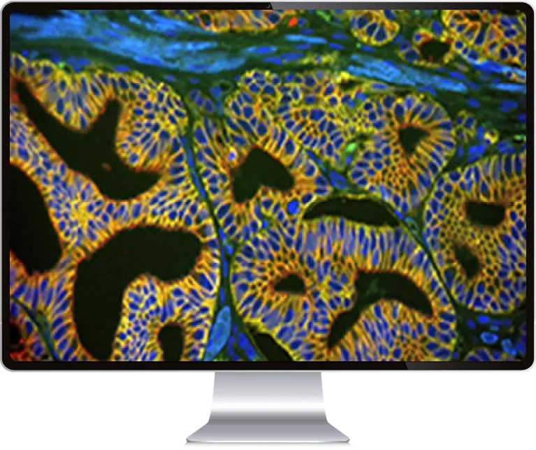
ASI’s HyperSpectral Imaging and Analysis system, provides an advanced multi-color solution for both brightfield and fluorescent samples, addressing research needs of clinical laboratories by extracting quantitative, spectral and morphological information on cell-biology, and providing molecular and cellular image insights.
Benefits to your lab
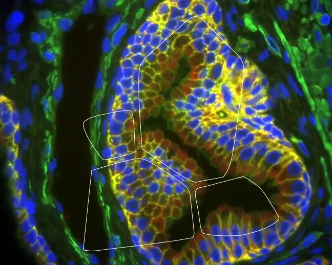
Uncover chemically similar areas hidden to the eye, create color-coded maps and compare the chemical makeup of components
Separate spectral
components to view
them as individual layers
Detect and classify objects, based on quantitative morphological and spectral content
Un-mix spectral components and remove background and auto-fluorescence for accurate, quantitative expression at every pixel
Unveil a world of research for a wide range of applications
- Cell biology
- Multicolor
- IHC Immunology
- Stem Cells
- Cell identification
- Bacteriology
- Infectious disease
- Pharmaceuticals
