

HiPath Pro
Ultimate Digital Pathology Workflow for Brightfield Images

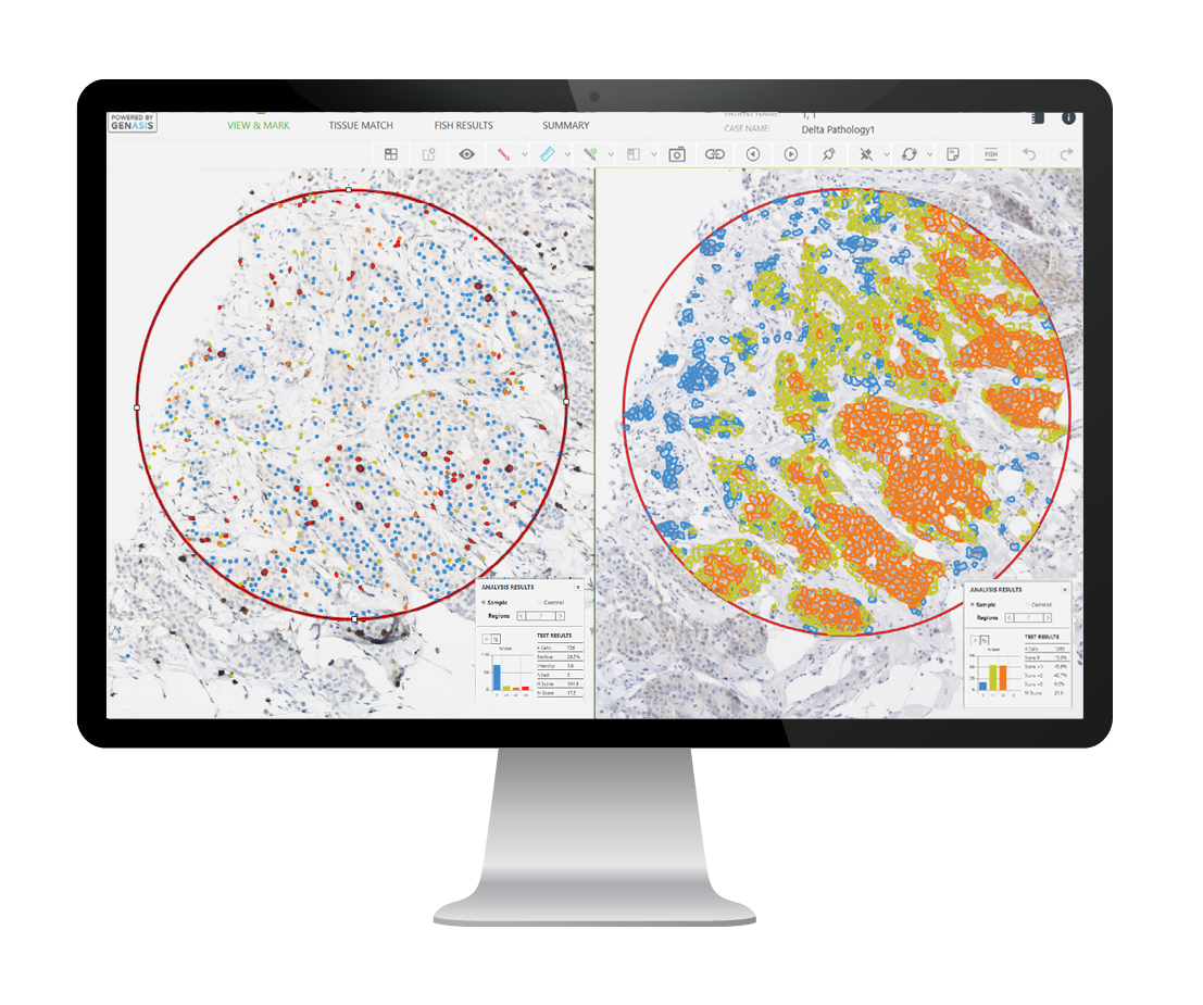

HiPath Pro
in a nutshell
HiPath Pro offers a range of advanced features and streamlined workflows for the scanning, visualization and analysis of brightfield pathology specimens.
Advanced marking and annotation tools, tissue matching across multiple stains, automated transfer of regions of interest and semi-quantitative IHC analysis are few of the tools offered by HiPath Pro, available for images acquired on the ASI scanning platform or third-party scanners.
Experience the unique capabilities of HiPath Pro to standardize results across your pathology practice.
Benefits to your lab
Intuitive whole slide
image navigation and
review up to 80X
digital zoom
Unique classifiers applicable to brightfield images from different formats
Semi-quantitative
IHC
analysis for ER, PR,
HER2, Ki67, P53,
PD-L1 and more
Analysis and scoring
of chromogenic
in situ hybridization
(CISH) stain
Standardize Your IHC Scoring with Computer Aided Analysis
HiPath Pro’s computer-aided IHC analysis module, powered by advanced algorithms, enables semi-quantitative scoring of tissue samples across an extensive variety of nuclear and membranous immunohistochemical stains. Applicable to images acquired on ASI and third-party scanners, HiPath Pro’s semi-quantitative IHC analysis serves as a valuable second opinion to enhance diagnostic confidence and reduce inter-observer variability.
